L56
1 /30Pages

L56
1 /30Pages
Extraits du catalogue

...going one step further
Ouvrir le catalogue en page 1
Latin A ilk-giving right breast and chest wall, front view, medially divided, representation of the M externally visible changes for breast gland inflammation (mastitis) on the inner half B Milk-giving right breast and chest wall, external half, cut surface C ilk-giving right breast and chest wall, inner half, cut surface, representation of breast gland M inflammation (mastitis) D Non-milk giving left breast and chest wall, front view, two sagittal cuts E on-milk-giving breast and chest wall; external half, external view, N skin windowed to illustrate the regional lymph nodes F Non-milk-giving...
Ouvrir le catalogue en page 2
Model of female breast L56 female breast and chest wall: milk-giving right breast with representation of an inflammation (mastitis) and non-milk-giving left breast with representation of various diseases The model consists of a milk-giving female right breast with the surrounding chest wall area and a nonmilk-giving female left breast with the surrounding chest wall area. Both parts of the model have a sagittal cut. The cut surfaces show the tissue of the breast gland as well as the deeper-lying anatomical structures such as the muscles, ribs, costal pleura, pulmonary pleura and lungs. A breast...
Ouvrir le catalogue en page 3
Model of female breast A ilk-giving right breast and chest wall, front view, medially divided, representation of the M externally visible changes for breast gland inflammation (mastitis) on the inner half B Milk-giving right breast and chest wall, external half, cut surface C ilk-giving right breast and chest wall, inner half, cut surface, representation of breast gland M inflammation (mastitis) D Non-milk giving left breast and chest wall, front view, two sagittal cuts E on-milk-giving breast and chest wall; external half, external view, N skin windowed to illustrate the regional lymph nodes...
Ouvrir le catalogue en page 4
Weibliches Brustmodell L56 Weibliche Brust und Brustwand: milchgebende rechte Brust mit Darstellung einer Entzündung (Mastitis), ruhende linke Brust mit Darstellung verschiedener Erkrankungen Das Modell besteht aus einer rechten milchgebenden weiblichen Brust mit dem umgebenden Brustwandbereich sowie einer linken ruhenden weiblichen Brust mit dem umgebenden Brustwandbereich. Beide Modellteile sind sagittal geschnitten. Die Schnittflächen zeigen neben dem Gewebe der Brustdrüse auch tiefer liegende anatomische Strukturen wie Muskeln, Rippen, Rippen- und Lungenfell und Lunge. An der rechten Brust...
Ouvrir le catalogue en page 5
Weibliches Brustmodell A ilchgebende rechte Brust und Brustwand, von vorne, median geschnitten, Darstellung M der außen sichtbaren Veränderungen bei Brustdrüsenentzündung (Mastitis) auf der inneren Hälfte B Milchgebende rechte Brust und Brustwand, äußere Hälfte, Schnittfläche C ilchgebende rechte Brust und Brustwand, innere Hälfte, Schnittfläche, Darstellung einer M Brustdrüsenentzündung (Mastitis) D Ruhende linke Brust und Brustwand, von vorne, zweifach sagittal geschnitten E uhende linke Brust und Brustwand, äußere Hälfte, von außen, Haut zur Darstellung der R regionären Lymphknoten gefenstert...
Ouvrir le catalogue en page 6
Modelo de mama de mujer L56 Mama de mujer y pared torácica: mama derecha lactante con representación de una inflamación (mastitis), mama izquierda en reposo con representación de distintas enfermedades El modelo se compone de una mama derecha lactante de mujer con región torácica circundante y una mama izquierda de mujer en reposo con región torácica circundante. El corte de ambos modelos es sagital. Las secciones de corte muestran, además del tejido de la glándula mamaria, estructuras anatómicas más profundas, como músculos, costillas, pleura y pleura pulmonar y pulmón. En la mama derecha se...
Ouvrir le catalogue en page 7
Modelo de mama de mujer A Mama lactante derecha y pared toracica, vista frontal, corte medial, representacion de los cambios externos visibles con inflamacion de glandula mamaria (mastitis) en la mitad interna B Mama lactante derecha y pared toracica, mitad externa, superficie de corte C Mama lactante derecha y pared toracica, mitad interna, superficie de corte, representacion de una inflamacion de la glandula mamaria (mastitis) D Mama izquierda en reposo y pared toracica, vista frontal, doble corte sagital E Mama izquierda en reposo y pared toracica; mitad externa, vista externa, piel con ventana...
Ouvrir le catalogue en page 8
Modèle de poitrine féminine Poitrine féminine et paroi thoracique L56 : poitrine droite donnant le sein avec représentation d’une inflammation (Mastitis), poitrine gauche au repos avec représentation de diverses affections Le modèle anatomique est constitué d’une poitrine droite féminine donnant le sein comprenant l’environnement de la paroi thoracique ainsi que d’une poitrine gauche au repos et de son environnement de la paroi thoracique. Les deux parties du modèle anatomique sont représentées en coupe Les surfaces de coupe montrent de profondes structures anatomiques parallèlement au tissu...
Ouvrir le catalogue en page 9
Modèle de poitrine féminine A oupe avant médiane de poitrine droite donnant le sein et paroi thoracique, représentation des C modifications externes notoires en cas d’inflammation des glandes mammaires (mastitis) de la face interne B Poitrine droite donnant le sein et paroi thoracique, moitié externe, surface de coupe C oitrine droite donnant le sein et paroi thoracique, moitié interne, surface de coupe, P représentation d’une inflammation des glandes mammaires (mastitis) D Poitrine gauche au repos et paroi thoracique, plan sagittal double, frontal E oitrine gauche au repos et paroi thoracique...
Ouvrir le catalogue en page 10
Modelo de Mama Feminina L56 Mama feminina e parede torácica: mama direita lactante com representação de inflamação (mastite), mama esquerda não lactante com representação de diversas doenças O modelo é composto de mama feminina direita lactante com a área da parede torácica adjacente, bem como de mama feminina esquerda não lactante com a área da parede torácica adjacente. Ambos os modelos são seccionados sagitalmente. Além dos tecidos da glândula mamária, as seções mostram também estruturas anatômicas mais profundas, como músculos, costelas, pleura parietal, pleura visceral e pulmões. A mama...
Ouvrir le catalogue en page 20Tous les catalogues et fiches techniques (PDF) 3B Scientific
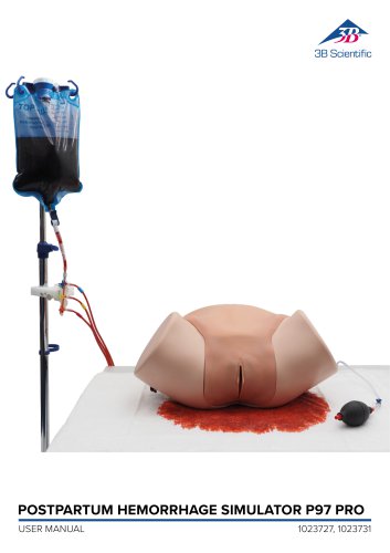
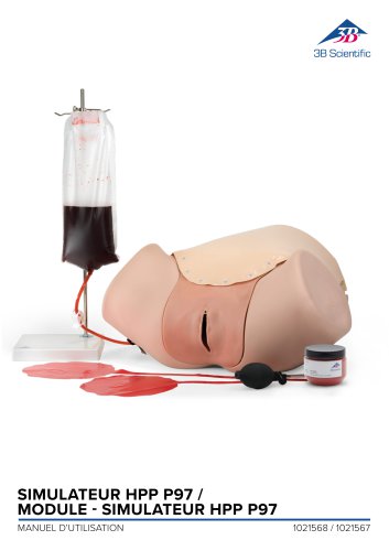 Simulateur HPP P97
14 Pages
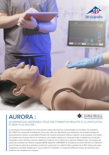 IngMar Sellsheet Aurora
2 Pages
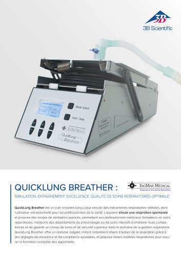 IngMar Sellsheet QuickLung
2 Pages
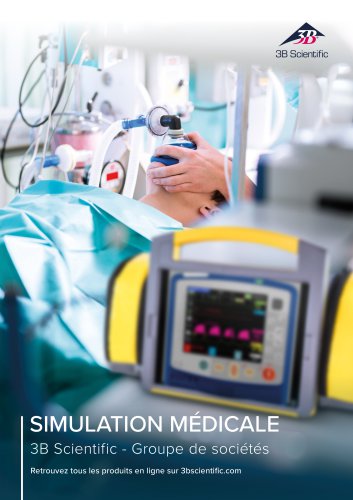 Medical Simulation
51 Pages
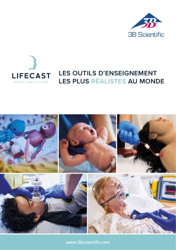 Lifecast Brochure
16 Pages
 Acupuncture
35 Pages
 P10/1,P11/1
11 Pages
 Catalogue Sciences Naturelles
196 Pages
 K17
K1716 Pages
 D25 Hémi-mandibule
16 Pages
 D20 Évolution de la dentition
12 Pages
 D10
D1012 Pages
 L50, L51, L55
36 Pages
 C15, C16, C17, C18, C20
9 Pages
 p80 SIMone Mode d' emploi
52 Pages
 P57 Mode d’emploi rapide
16 Pages
 N30 / N31 Mode d’emploi
12 Pages
 N15 Oreilles d’acupuncture
2 Pages
 P16 mode d'emloi
8 Pages
 poitrine féminine
30 Pages
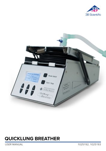 QuickLung Breather
12 Pages
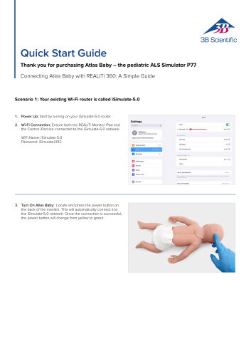 Quick Start Guide Atlas Baby
4 Pages
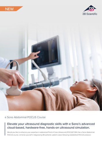 Sellsheet eSono Abdominal
3 Pages
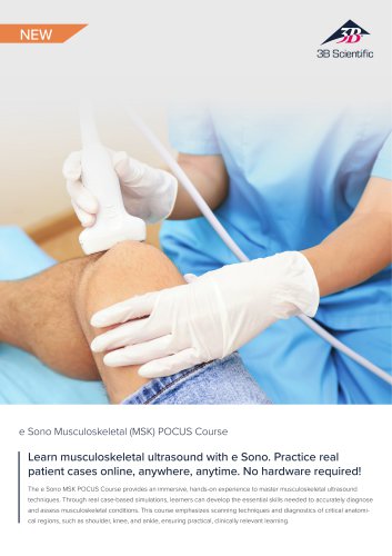 Sellsheet eSono MSK
3 Pages
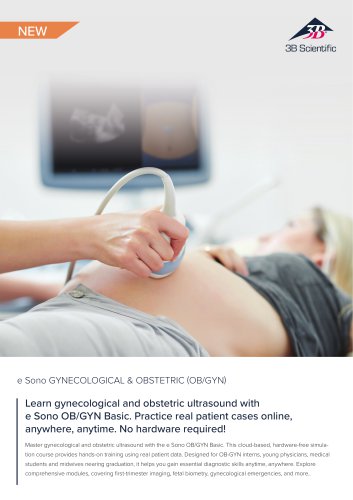 Sellsheet eSono OBGYN
3 Pages
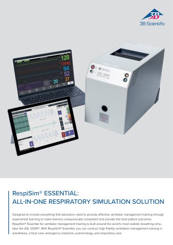 Sellsheet IngMar RespiSim
2 Pages
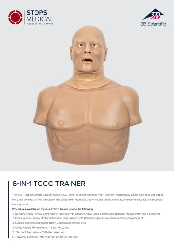 Sellsheet Stops 6N1 Trainer
2 Pages
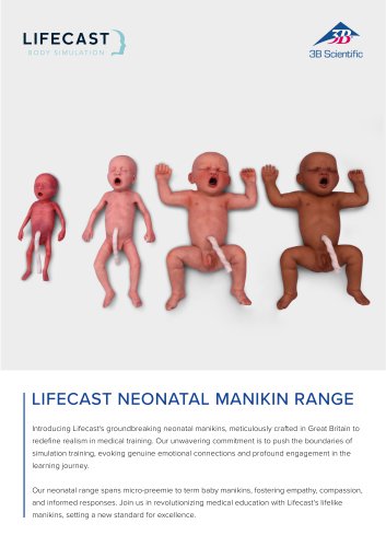 Sellsheet Lifecast Neonatal
3 Pages
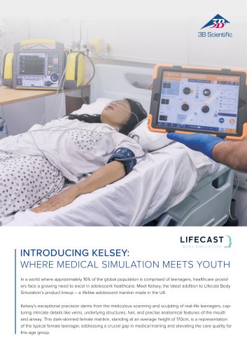 Sellsheet Lifecast Teenager
2 Pages
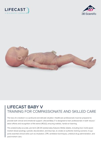 Sellsheet Lifecast Baby V
2 Pages
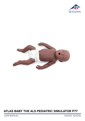 Product Manual Atlas Baby
16 Pages
 Sellsheet VSI 1025586
2 Pages
 Sellsheet VSI 1025528
2 Pages
 Sellsheet VSI 1025662
2 Pages
 Sellsheet VSI 1025616
2 Pages
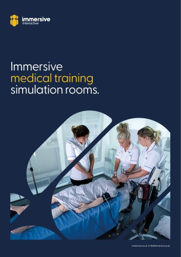 Immersive Brochure
13 Pages
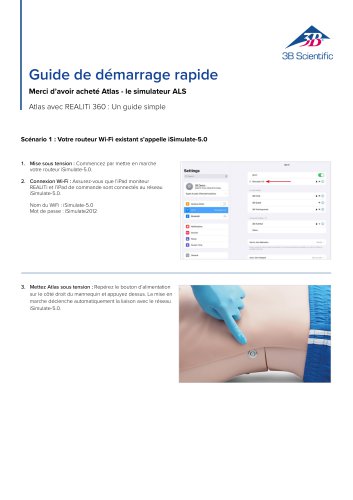
 Atlas Product Manual
18 Pages
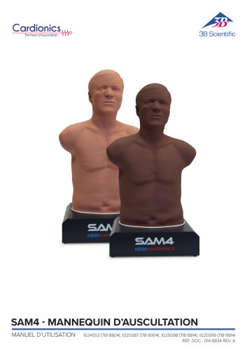
 Cardionics Brochure Simulation
23 Pages
 Best of Therapy
12 Pages
 Manual P120/P121/P122/P124/P125
60 Pages
 Medical Simulation EMS TCCC
9 Pages
 P10CCD product brochure
2 Pages
 P10CCD product manual
16 Pages
 P72+light Product manual
28 Pages
 P72+light Product brochure
2 Pages
 C41
C4116 Pages
 C18
C189 Pages
 G01
G0124 Pages
 3B Smart Anatomy
3 Pages
 M10
M1016 Pages
 A291
A29120 Pages
 F11
F1113 Pages
 P72
P7248 Pages
 B60
B6016 Pages
 A05/2 ,A11, A13
18 Pages
 A290 A291
20 Pages
 G21, G22
9 Pages
 K25
K2512 Pages
 K20, K21
12 Pages


![Notice d'utilisation - Bras pour injections I.V. P50-1 - P50/1 [1021418]](https://img.medicalexpo.fr/pdf/repository_me/67454/notice-utilisation-bras-injections-iv-p50-1-p50-1-1021418-249393_1mg.jpg)

![Notice d'utilisation - Bras d’entraînement au contrôle des hémorragies P102 - P102 [1022652]](https://img.medicalexpo.fr/pdf/repository_me/67454/notice-utilisation-bras-entrainement-controle-hemorragies-p102-p102-1022652-249357_1mg.jpg)
![Notice d'utilisation - Simulateur de soins des plaies et techniques de pansement - P100 [1020592]](https://img.medicalexpo.fr/pdf/repository_me/67454/notice-utilisation-simulateur-soins-plaies-techniques-pansement-p100-1020592-249351_1mg.jpg)

![Notice d'utilisation - Postpartum Hemorrhage Trainer - PPH Trainer P97 - P97 [1021568]](https://img.medicalexpo.fr/pdf/repository_me/67454/notice-utilisation-postpartum-hemorrhage-trainer-pph-trainer-p97-p97-1021568-249338_1mg.jpg)


![Notice d'utilisation - Estomac - 3B Smart Anatomy - K15 [1000302]](https://img.medicalexpo.fr/pdf/repository_me/67454/notice-utilisation-estomac-3b-smart-anatomy-k15-1000302-239673_1mg.jpg)


- Modèle anatomique 3B Scientific
- Modèle anatomique de formation 3B Scientific
- Modèle anatomique d'enseignement 3B Scientific
- Simulateur de formation 3B Scientific
- Simulateur pour soins généraux 3B Scientific
- Modèle anatomique de dents 3B Scientific
- Simulateur haut du corps 3B Scientific
- Modèle anatomique flexible 3B Scientific
- Modèle anatomique d'os 3B Scientific
- Modèle anatomique pour soins dentaires
- Modèle anatomique de crâne 3B Scientific
- Mannequin de formation 3B Scientific
- Simulateur de chirurgie 3B Scientific
- Stéthoscope médicale
- Modèle anatomique en plastique 3B Scientific
- Modèle anatomique de bouche 3B Scientific
- Modèle anatomique blanc
- Modèle vasculaire
- Simulateur patient
- Modèle vasculaire de formation