- Catalogues
- Olea Medical
- Medis suite CT
- Société
- Produits
- Catalogues
- News & Trends
- Salons
Medis suite CT
1 /14Pages

Medis suite CT
1 /14Pages
Extraits du catalogue

Imaging Solutions in a Heartbeat EÎMedis MEDICAL IMAGING NOS FONCTIONNALITÉS CLÉS • Analyse de la fonction myocardique • Analyse de la déformation par le strain pour LV, RV et Atria • Reformatage des données 3D en données 2D • Paramètres innovants tels que le déplacement vers l'intérieur • Rapide et facile à apprendre et à utiliser NOS PRINCIPALES CARACTÉRISTIQUES POUR LA RECHERCHE • Analyse CTA avec extraction automatique de l'arbre coronaire • Avec segmentation automatique des contours et de la lumière des vaisseaux • Rapport d'analyse de plaque par groupe
Ouvrir le catalogue en page 1
QUOI DE NEUF ? • Détection automatique de l'orientation des images long axe dans l'analyse des déformations • Ajout du Rubber banding et d'autres outils d'édition des contours pour en simplifier les changements dans l'analyses des déformations • Les résultats régionaux peuvent être exportés directement dans un fichier XML ou JSON. Cela s'applique au mouvement de la paroi et à toutes les valeurs de déformation régionales. Ces valeurs peuvent ainsi être automatiquement intégrées dans votre système de rapport ' Évaluation de la déformation longitudinale globale du ventricule gauche, sur les données...
Ouvrir le catalogue en page 2
Legal statement Medis Suite CT is based on image processing algorithms developed at the Division of Image Processing, Department of Radiology, Leiden University Medical Center, The Netherlands. Medis is a registered trademark of Medis Associated BV Medis Suite MRCT has market authorization in the EU, US, UK, Switzerland, Australia, Japan, Korea and Canada. Schuttersveld 9, 2316 XG Leiden 9360 Falls of Neuse Road, Suite 103 Medical Imaging P.O. Box 384, 2300 AJ Leiden, The Netherlands Raleigh, NC 27615-2484, USA Systems BV @Medis Medical Imaging in @Medis Medical Imaging www.medisimaging.com @MedisImaging...
Ouvrir le catalogue en page 3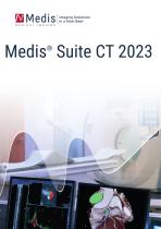
Imaging Solutions in a Heartbeat
Ouvrir le catalogue en page 4
• Auto-detection of papillary muscles and trabeculae with "MassK mode" • Quantification of EDV ESV, SV, %EF, CO, CI, indexed values (BSA and height), (time to) peak filling and ejection rate • Various BSA calculation methods for indexed results • Various normal ranges possible, calculation of z-scores M-MRM: QMASS REGIONAL FUNCTION MODULE • Analysis of regional parameters, such as wall motion, wall thickness, wall thickening and wall thickness changes over time • Regional results are part of the XML and JSON report output M-SMR: QSTRAIN CT • NEW: Automatic 2CH/3CH/4CH view recognition • NEW:...
Ouvrir le catalogue en page 5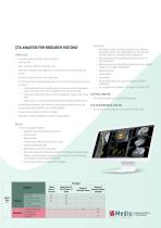
Data export: • All analysis results including coronary tree, contours, lesion parameters and vessel labels can be saved and reloaded again for reviewing and/or exporting • Easy data export for quantification data (Excel or co-py-to-clipboard) • Batch processing of quantified parameters from multiple studies into a single spreadsheet • Segment based • Lesion based • Slice based • Screenshots (jpeg, png, copy to clipboard, DICOM snapshots) • 3D visualization of plaque in 3D, export of lumen (.stl) CTA PVAT, ADD-ON • Peri-Vascular Adipose Tissue (PVAT) analysis CTA 3D WORKBENCH, ADD-ON • 3D visualization...
Ouvrir le catalogue en page 6
Imaging Solutions in a Heartbeat Legal Statement QMass and QFlow are based on image processing algorithms developed at the Division of Image Processing, Department of Radiology, Leiden University Medical Center, the Netherlands. Medis, QMass and QFlow are registered trademarks of Medis Associated BV. Medis Suite MRCT has market authorization in the EU, US, UK, Switzerland, Australia, Japan, Korea and Canada. Medis Medical Imaging Systems BV Schuttersveld 9, 2316 XG Leiden P.O. Box 384, 2300 AJ Leiden, The Netherlands P +31 71 522 32 44 F +31 71 521 56 17 E [email protected] Medis Medical...
Ouvrir le catalogue en page 8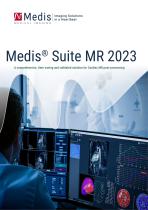
Imaging Solutions in a Heartbeat A comprehensive, time-saving and validated solution for Cardiac MR post-processing
Ouvrir le catalogue en page 9
M-MRM: QMASS REGIONAL FUNCTION MODULE (MR) • Analysis of regional parameters, such as wall motion, wall thickness, wall thickening and wall thickness changes over time • Regional results are part of the XML and JSON report output M-DCE: QMASS DELAYED SIGNAL INTENSITY (DSI) MODULE (INFARCT SIZE, T2W ANALYSIS, COMBINED DSI-T2W ANALYSIS) • Guided workflow for automatic infarct tissue quantification • Transfer contours from short axis cine stack • Various automated threshold calculation methods • Automatic infarct detection • Quantification of infarct size (% and mass), infarct transmurality • Quantifying...
Ouvrir le catalogue en page 10
M-TTM: QMASS T2/T2STAR ANALYSIS MODULE • Fast quantification of T2* decay time and decay rate • Color overlay • Correct for breathing motion M-TOM: QMASS T1 ANALYSIS MODULE • Measure T1 value based on automatic motion corrected T1 Maps • Calculation of T1 relaxation time in MOLLI and Look Locker sequences • Calculation of residual maps • Automatic Motion Correction • Color overlay • Correction for breathing motion M-FLX: QFLOW APP • Phase-contrast MR blood flow analysis • Automatic contour detection • Copy of contours in forward and backward diretion • Various background correction methods to...
Ouvrir le catalogue en page 11
M-HDF: QSTRAIN HEMODYNAMIC FORCES ADD-ON, FOR RESEARCH MS-REL: QMAP T1&T2 RELAXOMETRY, FOR USE ONLY RESEARCH USE ONLY • Create parametric maps for Tl, Tl*, T2 and T2* • Supports LL, MOLLI, SR, T2 prep and console generated maps • Correction factor • Offset, scaling, fit residual error • Display of relaxation graphs • Flexible manual motion correction • Flexible co-registration of T1 native (pre-contrast) and T1 post-contrast maps • Comprehensive results for myocardial segments and up to 4 ROI's and segments • AHA 16 segment model results and bull's eyes • Save maps as DICOM • Save results to...
Ouvrir le catalogue en page 12Tous les catalogues et fiches techniques (PDF) Olea Medical
 Breast in Cloud
12 Pages
 Automation Platform Brochure FR
18 Pages
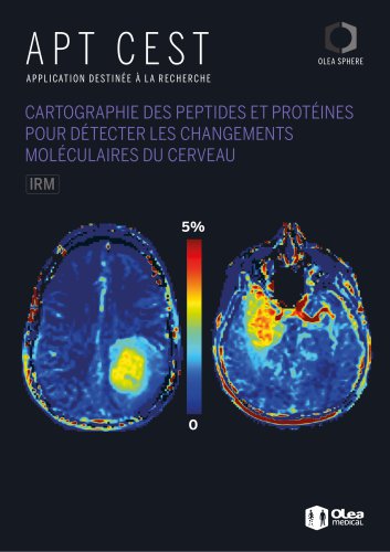 APT CEST
4 Pages


- Logiciel de visualisation
- Logiciel pour imagerie médicale
- Logiciel Windows
- Logiciel automatisé
- Logiciel de traitement
- Logiciel de traçabilité
- Logiciel de mesure
- Logiciel de chirurgie
- Logiciel d'importation
- Logiciel serveur
- Logiciel sur site
- Logiciel d'analyse de données
- Logiciel pour cardiologie
- Logiciel TDM
- Logiciel d'évaluation
- Logiciel d'analyse d'image
- Logiciel d'interprétation
- Logiciel de dépistage
- Logiciel de navigation
- Logiciel pour communication